How to put a DSLR camera on a microscope
I know that for a lot of you, microscope photography is second nature, but being the astrophotographer, I'm working at the other end of the spectrum and this is something that's new to me.
We sell a range of microscopes. They start as very basic microscopes - essentially for kids and students, and go up to what I might call a middle-range professional instrument, suitable for engineering laboratories, vets, botanists, pest controllers, etc. Nothing like cutting-edge university labs or electron microscopes, sorry.
We've also got a number of ways of getting an image from a microscope. We'd prefer to use a separate imager, which is essentially a camera sensor that slides into the eyepiece holder or trinocular head.
We've even got a few entirely digital microscopes, both hand-held or with a screen on a microscope body. If pushed, we can even get a mobile phone camera to the eyepiece.
But the other day, a customer asked about how to attach a DSLR to a microscope. I've never really considered putting something as large as a DSLR onto one.
So I decided to figure out how it's done.
What you will need
It all comes down to the humble t-thread. This is an industry-standard M42x0.75mm thread, and it's used to connect all manner of diverse things. Going through this thread, you can connect DSLRs, CMOS cameras, filters, eyepiece projectors, etc. onto telescopes, spotting scopes, filter wheels, and, as it turns out, microscopes.
In this case, the adapters I needed were my own well-worn adapter for a Pentax K mount, and the saxon microscope adapter.
t-ring
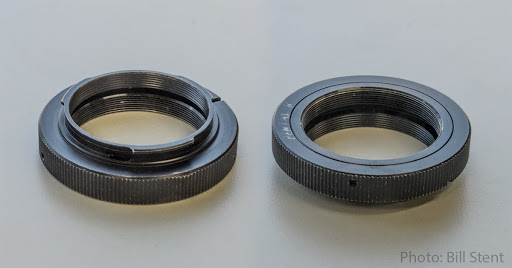
From the camera side, you need an adapter that fits onto your DSLR's bayonet mount. When you take your lens off, this little jigger, which is known as a t-ring, goes onto the camera body. Which one you need depends on your camera, of course, but they all end in a female t-thread.
From the microscope side, you need a second adapter that slides into the eyepiece holder or trinocular head. This adapter ends in a male t-thread.
 |
| A t-ring for a Pentax DSLR |
Here's the t-ring. Many readers will already be well familiar with this type of adapter - this one has a Pentax K-mount bayonet on it, because that's the type of DSLR I use. Of course, if your camera is a Canon or a Nikon or some other brand, you need the t-ring that matches your camera. On the other end of the t-ring is the all-important female t-thread.
Microscope adapter
And here's the microscope adapter. The male t-thread is on the bottom, and this threads onto the t-ring. As you can see, the top end is simply a straight cylinder of about 23.2mm that slides into whatever part of the microscope you're going to use.What you can do with them
I had a fun half hour or so, trying my camera and the adapters out on three of the microscopes we have on display in the shop. I could have spent more time on it - to be honest, it was quite a lot of fun.
We've got two major types of microscopes on display, dissecting and biological, and I took photos through both types.
A bit about microscope types
A dissecting type normally has two eyepieces so you can get 3-D views of your subject. This way, you can move the specimen about and manipulate it while getting close-up views of what you're working on. Because it's meant to be used for large items, like flowers, rocks or even frogs, it has low(ish) magnification.
The other major type of microscope is for biological work. Because the objective lens can get much closer, magnification is much higher, often up to 1600 times. You use a biological microscope for looking at things like plant or animal cells, or tiny things like insect parts - anything you can get onto a microscope slide, really.
saxon RST Researcher NM11-2000 dissecting microscope
 |
saxon RST Researcher NM11-2000 |
This microscope has a "trinocular" head - that is, not only are there two eyepieces, there's a third place for the camera, which I used. I found an old circuit board for my photos on this microscope.
Here's the photo I got. This was the full frame in my DSLR. I had to muck about with the exposure a bit. The light is from the side in this photo, but you can see there's a very slight touch of darkening in the corners. As is often the case, low magnification instruments are very easy to use, with good bright images and wide fields of view.
Here's a detail from that photo - I've attempted to show you as close to a one-pixel in the camera to one-pixel on your screen as I can, but this is going to depend on how you're displaying this article.
I'm quite pleased with this photo. Such a setup could be quite useful for some professions - and a lot of fun for some kids.
saxon RBT Researcher Compact biological microscope
 |
| saxon RBT Researcher Compact |
The saxon RBT Researcher Compact is a high-magnification stereo microscope. It also has a trinocular head, so the camera went there. I have taken better photos in my time, but because my DSLR was part of the photo I had to use my phone!
This one wasn't quite as easy to use as the dissecting microscope. I had to take a few test shots in manual mode before I was happy with the lighting. You can also see that the image was smaller than the size of the camera sensor, resulting in a dark shadow around the outside. This is called vignetting, and it is a bit of a waste of pixels. I could also have spent more time on fine focusing.
The full-size image is quite good, with the larger cells clearly visible. I'm sure a bit of practice would make this a very useful image.
saxon SBT ScienceSmart biological microscope
 |
| saxon SBM ScienceSmart |
Finally, this second biological microscope was a much cheaper saxon SBM ScienceSmart. This is a microscope with lower magnification. It also has a single eyepiece, so I inserted the camera into the eyepiece holder. The phone photo shows it looks a little ungainly, but it was quite stable. The eyepiece turret on this microscope can also rotate, meaning I could have had the camera on the other side, but I wanted to see the back of the camera.
This is the whole frame photo. I was very pleased with the image I got, even though it showed a little vignetting. It's smaller than the Researcher Compact partly because of the lower magnification of the microscope.If nothing else, it shows that the slides we use for our display models need to be cleaned!

The full-size photo shows the cells in detail. I note a little softening of focus in some areas of the field, which may be a result of dirt in the optics, and not necessarily optical quality. These microscopes have been out on display for several years!
Conclusion
Overall, I'm pretty pleased with these photos. They were a lot easier to get than I'd expected. I'm reasonably used to photographing things through optical equipment, but the fact that I got these photos in about a half hour shows it's not difficult for you to do.
The ability to take high resolution DSLR photos through a microscope is useful for a lot of people, and fun for others.


Comments
Post a Comment